Why Microscope-Enhanced Endodontics?
Root canal anatomy is extraordinarily complex and microscopic. Canals can be finer than a human hair, calcified, curved, or multi-branched. Using our Zeiss Surgical Operating Microscope, Dr. Ken Heritage achieves up to 20x magnification with direct coaxial illumination.
This ensures that hidden extra canals (such as MB2 in upper molars), subtle fractures, calcifications, and anatomical anomalies are clearly visualized, disinfected, and sealed hermetically.
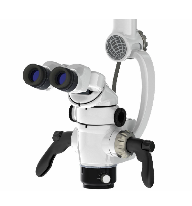
Zeiss High-Power Operating Microscope
Equipped with specialized optics to identify minute canal entries, evaluate pulp floor anatomy, and remove fractured instruments without removing healthy tooth structure.
Comprehensive Endodontic Treatments Provided:
Initial Primary Root Canal Treatment
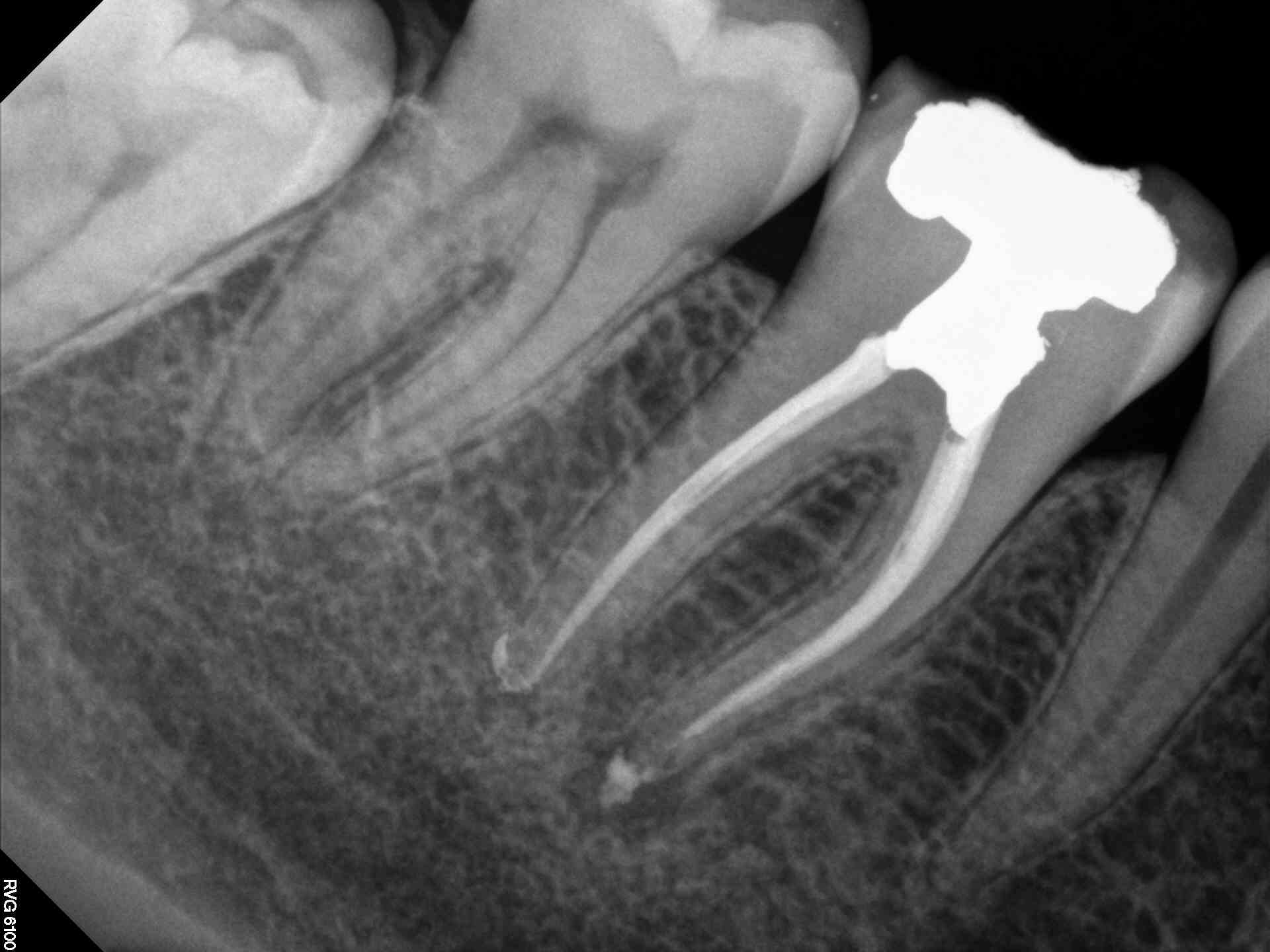
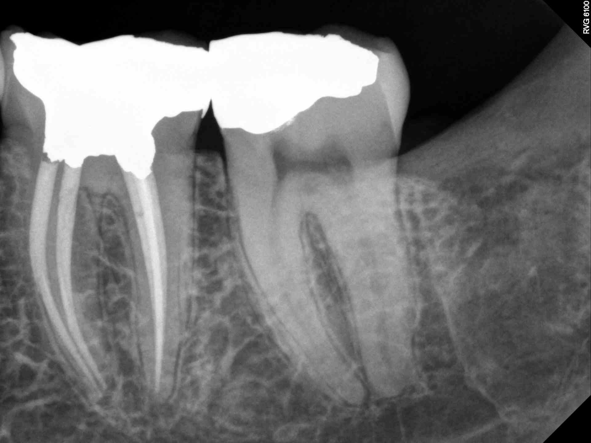
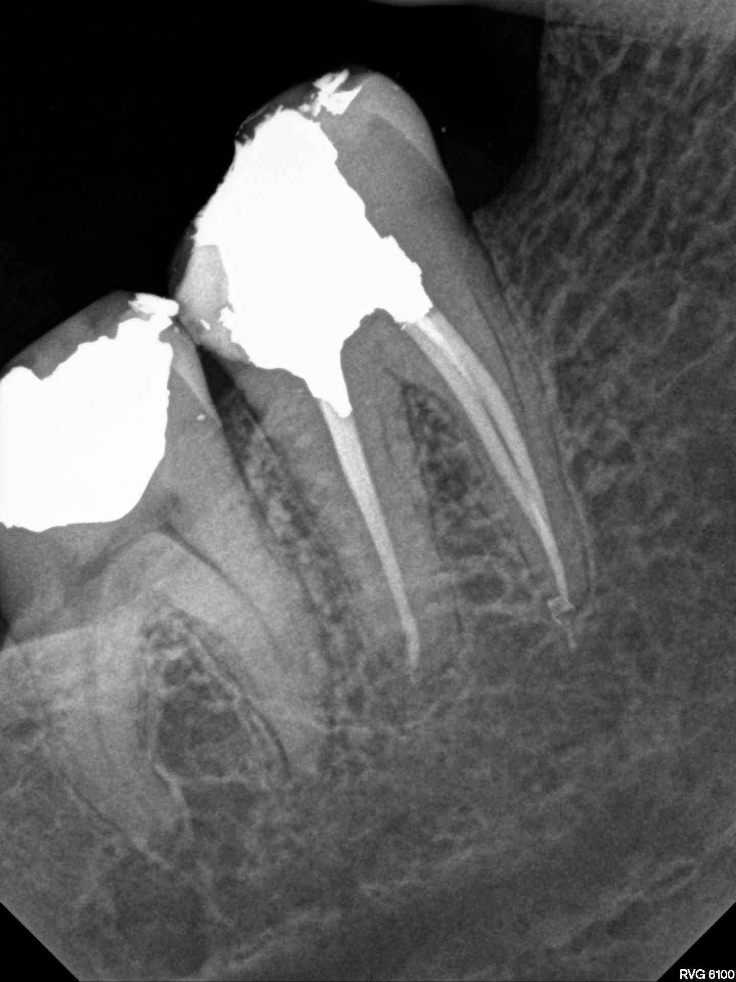
Thorough mechanical shaping using flexible Nickel-Titanium (NiTi) rotary instruments, comprehensive ultrasonic irrigation to eliminate bacterial biofilms, and three-dimensional obturation.
Endodontic Retreatment (Failed Previous Procedures)
When an existing root canal fails to heal due to persistent infection, missed canals, or poor coronal seal, we safely remove old filling materials, re-disinfect the complex root spaces, and re-seal.
Removal of Broken Instruments & Metal Posts
Ultrasonically dislodging and retrieving separated instruments, silver points, or cast posts under direct microscopic vision without compromising root strength.
Microsurgical Apicoectomy (Root-End Surgery)
When non-surgical treatment is impractical, microsurgery allows surgical resection of the infected root apex, ultrasonic cavity preparation, and placement of bioceramic MTA root-end seals.
Advanced 3D Obturation: System B Heat Condensation
To prevent re-infection, clean root canals must be filled in three dimensions. We utilize the System B continuous wave heat condensation technique (pioneered by Dr. L. Stephen Buchanan). This warms biocompatible gutta-percha, allowing it to flow seamlessly into lateral canals, accessory deltas, and irregularities.